The Occipital Place Area: Your Brain's Guide to Moving Through Space
Three scene-selective brain regions divide the work of scene perception — the OPA, named in 2013 after TMS evidence gave it a causal role, handles the local elements of visual scenes and the boundary information your brain needs to navigate through immediately visible space. This article covers its anatomy, the fractured-rooms experiment that revealed its local processing bias, the dynamic scenes result linking it to first-person locomotion, and the virtual-reality boundary navigation study that placed it at the perceptual origin of the spatial navigation circuit.

You can walk through a doorway, sidestep a chair, and turn left at the end of a corridor without thinking about any of it. What makes that possible isn't one system but three distinct cortical regions that carve up the problem of "knowing where you are" into specialized sub-problems. Two of them — the parahippocampal place area (PPA) and the retrosplenial complex (RSC) — have been studied intensively since the late 1990s. The third, the occipital place area (OPA), spent its first decade as little more than a footnote, often known only by its anatomical address: the transverse occipital sulcus, or TOS. That changed in 2013, when a TMS study gave the region its name and its first causal role in scene perception.
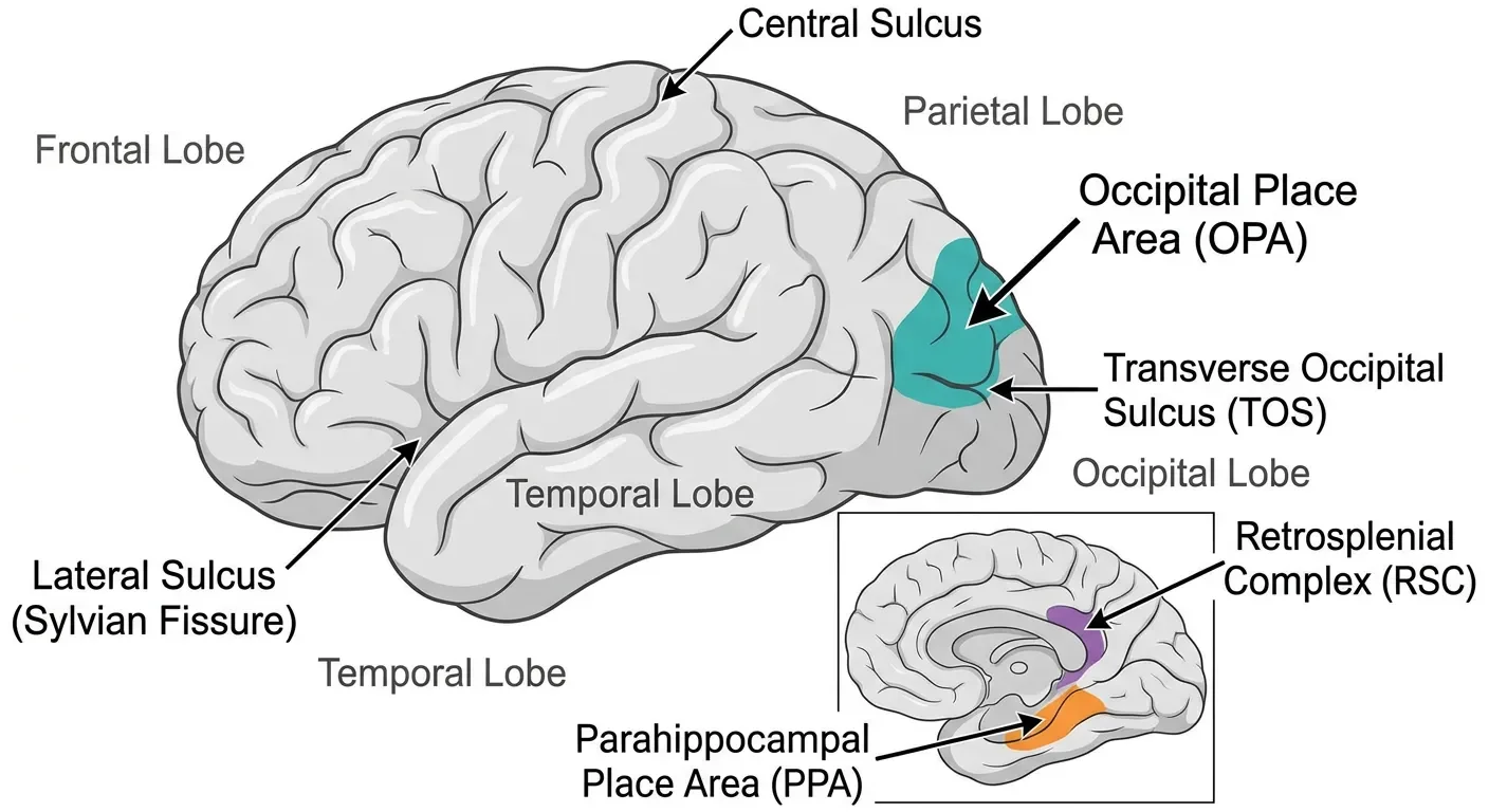
Where it lives and what it responds to
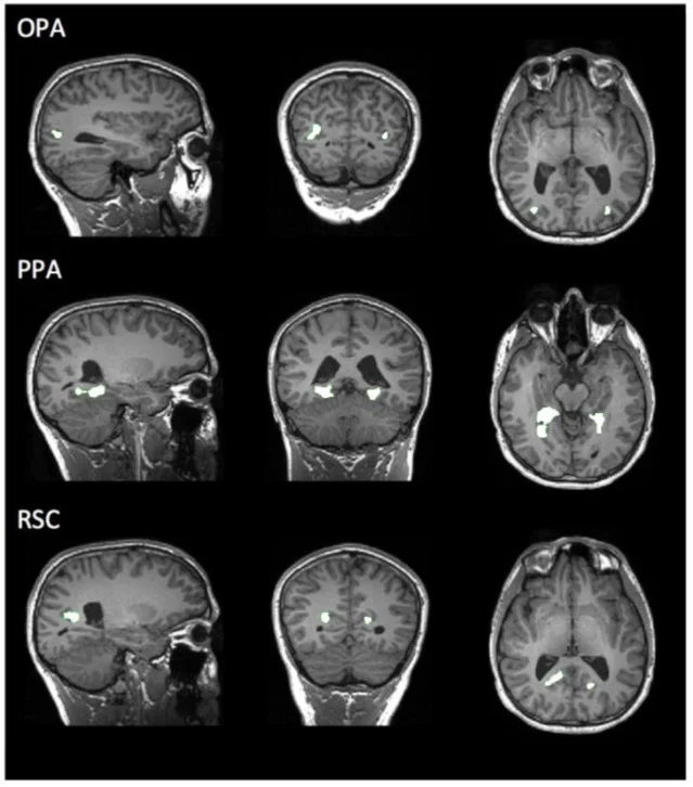
The OPA sits at the junction of the occipital and parietal lobes, clustered around the transverse occipital sulcus in posterior cortex. 1 It is scene-selective in the standard fMRI sense: it responds more strongly to photographs of places, rooms, streets, and landscapes than to faces, objects, or scrambled images. In this respect it looks like the PPA and RSC. But its anatomical position is meaningfully different from those two: the PPA is tucked into the parahippocampal gyrus on the ventral temporal surface, and the RSC sits near the posterior cingulate in medial cortex. The OPA is lateral and posterior — the most occipital of the three, physically close to early visual processing regions.
The naming paper: giving TOS a better name
For roughly a decade the region was called "TOS" after its anatomical neighbor the transverse occipital sulcus — a purely geographic label that said nothing about function. The name change to "occipital place area" came from Dilks, Julian, Paunov, and Kanwisher at MIT in a 2013 Journal of Neuroscience paper whose headline result was also its methodological anchor: TMS applied to the right OPA disrupts scene perception selectively. 1
The study ran two experiments. In the first, participants saw a scene (or a face), then had to discriminate a fine-grained morph of that stimulus from a slightly different one — a staircase procedure tuned to measure the minimum perceivable difference. TMS to the right OPA raised the discrimination threshold for scenes but not faces. TMS to the right occipital face area (OFA) did the reverse — impaired faces but not scenes. Each region broke only its own category.
In the second experiment, participants had to categorize scenes (beach, city, kitchen, forest) and objects (camera, chair, car, shoes) at threshold presentation durations. Again, TMS to OPA hurt scene categorization but not object categorization; TMS to the right lateral occipital complex (LOC) hurt objects but not scenes. The double dissociation across both experiments ruled out a generic "disruption" account and confirmed that the OPA is a causal, category-specific component of the scene-processing circuit.
Dilks and colleagues renamed the region OPA rather than TOS for three reasons: TMS evidence strengthened its functional identity as a place/scene area; the region is functionally rather than anatomically defined; and crucially, in some participants the peak activation falls outside the transverse occipital sulcus proper. 1
Local elements, not global layout
Once the OPA had a name and a causal role, the next question was what, specifically, it extracts from a scene. A 2016 NeuroImage paper by Kamps, Julian, Kubilius, Kanwisher, and Dilks provided a clear answer: the OPA analyzes local scene elements, not global spatial structure. 2
The key manipulation was "fractured rooms." Take a photograph of a bare apartment room, chop it into its wall, floor, and ceiling surfaces, then scramble their arrangement so the space no longer forms a coherent enclosure. The global layout is destroyed; the local surface elements — the individual planes — are all still present. When participants viewed intact versus fractured rooms, PPA and RSC responded significantly more to intact rooms (the coherent global layout mattered to them). The OPA showed no significant difference between intact and fractured conditions. It was equally satisfied by the scattered surfaces as by the properly assembled room.
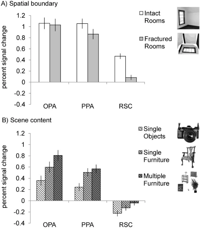
A second manipulation tested scene content. Participants viewed images containing a single non-furniture object, a single piece of furniture, or multiple pieces of furniture. The OPA's signal increased with furniture count; the PPA's did not vary significantly with the number of items. The pattern across both manipulations fits a clean story: OPA tallies local elements — surfaces, objects — while PPA and RSC compute the global properties of the environment.
This division maps onto an analogy from the face and body systems covered in earlier articles in this series. Just as the OFA detects local face parts (eyes, nose, mouth) regardless of their configuration while the FFA requires a properly assembled face, the OPA detects local scene elements regardless of global arrangement while the PPA requires a coherent spatial layout.
Visually guided navigation: the first-person view
What does analyzing local elements buy you functionally? A 2016 Cortex paper by Kamps, Lall, and Dilks proposed a concrete answer: first-person perspective motion through the immediately visible environment. 3
The experiment compared fMRI responses to two conditions: "Dynamic Scenes" (3-second video clips filmed by walking through a space at eye level, so the participant experienced the actual visual flow of locomotion) and "Static Scenes" (still frames taken from the same clips but presented in shuffled order, removing any sense of forward motion). If the OPA cares about navigating through the visible scene rather than just recognizing it, it should respond more to the dynamic version.
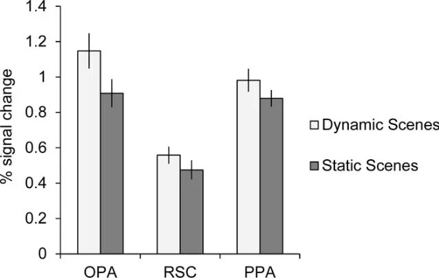
OPA showed a significantly greater response to Dynamic than Static Scenes, and this difference was larger than the corresponding difference in RSC or PPA. Two controls ruled out simpler explanations. First, the dynamic advantage in OPA was not present for faces filmed in motion versus static face images — so it isn't generic motion sensitivity. Second, the advantage was larger than in MT, a domain-general motion region, ruling out the possibility that OPA simply inherited more low-level motion signal from early visual cortex.
The RSC didn't show the same dynamic preference. That's theoretically important. RSC is thought to support broader environmental knowledge — knowing where a location sits in a larger cognitive map, orienting by distant landmarks. The OPA, by contrast, looks like it handles the immediate local scene during active passage through it. The two systems appear to serve visually guided navigation (OPA, the local view) versus map-based navigation (RSC, the wider spatial context).
Boundaries: what walls do that floors don't
The most direct test of OPA's role in navigation came in a 2016 Current Biology study by Julian, Ryan, Hamilton, and Epstein at the University of Pennsylvania. 4 The experiment used virtual reality.
Participants learned the locations of objects inside a circular virtual arena that contained both a distinctive wall boundary and a movable landmark object. On each trial they navigated to a remembered location and received feedback. Critically, between blocks the landmark was moved relative to the wall — so objects either stayed fixed to the boundary (boundary-tethered objects) or stayed fixed to the landmark (landmark-tethered objects). Before each block, continuous theta-burst TMS was applied to either the right OPA or a vertex control site.
The result was a sharp dissociation: TMS to the OPA significantly impaired accuracy for boundary-tethered objects but had no effect on landmark-tethered objects. When the OPA was disrupted, participants shifted toward using the landmark even for boundary-coded locations — the system that normally anchors position to wall geometry had been knocked offline.
A follow-up experiment clarified what kind of boundary matters. When the arena edge was defined by a floor marking rather than a wall, OPA stimulation no longer impaired navigation. The OPA appears to be specifically sensitive to vertical boundary surfaces — the kind that obstruct movement and define the outer shape of navigable space — rather than just large visual features generally.
This finding ties the OPA into a circuit that stretches from early visual perception all the way to hippocampal place cells. Behavioral and neural work going back decades shows that animals (and humans) code locations preferentially by their relationship to environmental boundaries, and that boundary-sensitive cells exist in the hippocampus and subiculum. 4 The Julian et al. result suggests that the OPA is where visual input to that system originates — the perceptual stage that extracts wall boundaries and feeds them downstream to memory structures.
A three-region framework for scene processing
By 2019, Epstein and Baker had synthesized the evidence into a clear three-part framework. 5 Each of the three scene-selective regions has a characteristic functional signature:
| Region | Location | Primary function |
|---|---|---|
| OPA | Lateral posterior occipital cortex | Local scene elements; visually guided navigation; boundary perception |
| PPA | Parahippocampal gyrus (ventral temporal) | Global spatial layout; place recognition; environmental context |
| RSC | Posterior cingulate / retrosplenial cortex (medial) | Landmark-based navigation; spatial memory; map-level orientation |
The OPA handles the immediate visual environment — what's right in front of you and how to move through it. The PPA identifies the kind of place and its broad spatial structure. The RSC situates that place within a larger mental map. You need all three to walk confidently through an unfamiliar building.
Open questions
Several things about the OPA remain genuinely unsettled. The TMS evidence establishes a causal role in perception, but it cannot cleanly separate encoding from retrieval: disrupting the OPA during a spatial memory task could impair the initial extraction of boundary information, the retrieval of that information later, or both.
A second open question is the OPA's connectivity. Functional imaging suggests it is linked to both the PPA and to posterior parietal regions involved in spatial attention and action planning. 4 Whether information from the OPA reaches the hippocampus primarily via the PPA route, via a parietal route to the RSC, or both is not settled.
Third, the OPA develops relatively late. Studies with children show that OPA's scene selectivity is adult-like by age 7 or 8, but its sensitivity to left-right "sense" information in scenes lags behind — 8-year-olds show it but 5-year-olds typically don't. 6 This developmental pattern suggests that the region's selectivity for navigationally-relevant spatial properties is experience-dependent in a way that basic scene selectivity is not.
Landmark paper: Dilks, D.D., Julian, J.B., Paunov, A.M., & Kanwisher, N. (2013). The occipital place area is causally and selectively involved in scene perception. Journal of Neuroscience, 33(4), 1331–1336. https://doi.org/10.1523/JNEUROSCI.4081-12.2013 1
Course connection: MIT 9.13 "The Human Brain" (Prof. Nancy Kanwisher) covers the OPA in the context of scene-selective cortex and functional imaging methods for identifying category-selective regions. The OPA connects directly to lectures on the parahippocampal place area, the fROI method, and the question of how different scene regions contribute to navigation — topics covered in this series on the PPA and the fROI method.
Añade más opiniones o contexto en torno a este contenido.